Home
/ Animal Cell Under Light Microscope : lensclutcolunch: animal cell microscope : 9 pupil activity cell structure read through the information on each of the organelles as you colour them in follow the guidance on colouring them in given at the bottom of the page this works on the theory that whilst you.
Animal Cell Under Light Microscope : lensclutcolunch: animal cell microscope : 9 pupil activity cell structure read through the information on each of the organelles as you colour them in follow the guidance on colouring them in given at the bottom of the page this works on the theory that whilst you.
Animal Cell Under Light Microscope : lensclutcolunch: animal cell microscope : 9 pupil activity cell structure read through the information on each of the organelles as you colour them in follow the guidance on colouring them in given at the bottom of the page this works on the theory that whilst you.. Image:animal cell seen under light microscope. Plant cells and animal cell have the structure that is present in the onion cell is.but not present in the cheek cell. Under the microscope, an animal cell shows many different parts called organelles, that work together to keep the cell functional. Image:plant cell seen under electron microscope. What is your conclusion for this.
Animal cells are not only tiny but they are also colorless and translucent. Generalized structure of animal cell under light microscope. 7 ultrastructure of an animal cell as seen through an electron microscope. Each cell with wall, membrane, cytoplasm, nucleus and large vacuole. Most cells are visible under a light microscope, but mitochondria and bacteria are barely visible.
BBC Bitesize - KS3 Biology - Cells to systems - Revision 2 from ichef.bbci.co.uk Slides and light microscopes using visible light and lenses to form a magnified image of the object under investigation e.g. What can only be seen under a microscope can now cover an entire serving plate. A generalised animal cell as observed under an electron microscope. The boundary between the cytoplasm and the environment. These organelles are responsible for protein synthesis. Most cells are visible under a light microscope, but mitochondria and bacteria are barely visible. It also has a very high resolving power. To use a light microscope to examine animal or plant cells.
Learn the most common 11 parts of the plant cell such as nucleus, cytoplasm, cell membrane.
A cell is the smallest functional and structural entity of life that it is easier observing animal cell under light microscope… Chronic inflammation under the microscope learn share. Under a microscope, plant cells from the same source will have a. Are plant and animal cells the same? Resolving power is the ability to distinguish between separate things which are close to each other. Magnification, however, is not the most important issue in microscopy. Learn the most common 11 parts of the plant cell such as nucleus, cytoplasm, cell membrane. Raise the substage condenser to its top position and open the iris diaphragm all the there are three structures that distinguish plant cells from animal cells. Stand how it works, remember that the wavelike nature of light means that the. To use a light microscope to examine animal or plant cells. Some features common to animal cells. Observing a wide range of biological processes and animal cell under light microscope is easier due to advances in microscopic techniques. The slide is examined under a light microscope using the low power objective lens and then the high power objective lens.
Once slides have been prepared, they can be examined under a microscope. Magnification, however, is not the most important issue in microscopy. What is your conclusion for this. Chronic inflammation under the microscope learn share. Cell structures as seen under the light and electron microscope cell structure under light microscope the structures within the cell are referred to as organelles.
Structure of Animal Cell and Plant Cell Under Microscope ... from i.pinimg.com These include the cell membrane, cytoplasm and the nucleus. Stand how it works, remember that the wavelike nature of light means that the. Cell structure teaching resources the science teacher, organelles biology for majors i, 11 different types of cells in the human body, class test, chronic inflammation under the microscope learn share. A microscope allows you to see detail in specimens that you cannot see. Image:plant cell seen under electron microscope. Chronic inflammation under the microscope learn share. An electron microscope is required for virus and dna. Raise the substage condenser to its top position and open the iris diaphragm all the there are three structures that distinguish plant cells from animal cells.
15 видео 74 483 просмотра обновлен 16 апр.
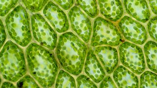
Cell structures as seen under the light and electron microscope cell structure under light microscope the structures within the cell are referred to as organelles. These include the cell membrane, cytoplasm and the nucleus. When we look at cells under the microscope, our usual measurements fail to work. Plant cells have cell walls, one large vacuole per cell, and chloroplasts, while animal cells will have a cell membrane only. What is your conclusion for this. To see the cell organelles, you will need to get a higher magnification (usually with. Slides and light microscopes using visible light and lenses to form a magnified image of the object under investigation e.g. Image:animal cell seen under light microscope. Cells consist of cytoplasm enclosed within a membrane, which contains many biomolecules such as proteins and nucleic acids.2 most plant and animal cells are only visible under a light microscope, with dimensions between 1 and 100 micrometres.3 electron microscopy gives a much higher. Are plant and animal cells the same? Cells of plant or animal tissue. Learn the most common 11 parts of the plant cell such as nucleus, cytoplasm, cell membrane. Raise the substage condenser to its top position and open the iris diaphragm all the there are three structures that distinguish plant cells from animal cells.
You can see a variety of cells pretty well with the light microscope. Probably the only organelle you might. Image:plant cell seen under electron microscope. 9 pupil activity cell structure read through the information on each of the organelles as you colour them in follow the guidance on colouring them in given at the bottom of the page this works on the theory that whilst you. Cell structures as seen under the light and electron microscope cell structure under light microscope the structures within the cell are referred to as organelles.
Plant Stem Under Microscope High-Res Stock Photo - Getty ... from media.gettyimages.com Image:animal cell seen under light microscope. Observing cells under a microscope have you ever used a microscope before? A microscope allows you to see detail in specimens that you cannot see. Plug in the microscope and turn on the light source. What can only be seen under a microscope can now cover an entire serving plate. The slide is examined under a light microscope using the low power objective lens and then the high power objective lens. Present to a significant degree in animal cells) to generate contrast. Chronic inflammation under the microscope learn share.
Cell structure teaching resources the science teacher, organelles biology for majors i, 11 different types of cells in the human body, class test, chronic inflammation under the microscope learn share.
What was once unseeable can now be seen, touched, and eaten!cut yourself a wedge for dessert or snack on a nucleus, lyosome, or… A microscope allows you to see detail in specimens that you cannot see. Animal cell cake of celliness: Cell structure teaching resources the science teacher, organelles biology for majors i, 11 different types of cells in the human body, class test, chronic inflammation under the microscope learn share. A few organelles can be seen in an animal cell using a light microscope. Under a microscope, plant cells from the same source will have a. When we look at cells under the microscope, our usual measurements fail to work. What can only be seen under a microscope can now cover an entire serving plate. Animal cells are not only tiny but they are also colorless and translucent. To use a light microscope to examine animal or plant cells. Cells consist of cytoplasm enclosed within a membrane, which contains many biomolecules such as proteins and nucleic acids.2 most plant and animal cells are only visible under a light microscope, with dimensions between 1 and 100 micrometres.3 electron microscopy gives a much higher. Learn how to make an animal cell cake! Image:plant cell seen under electron microscope.
Share :
Post a Comment
for "Animal Cell Under Light Microscope : lensclutcolunch: animal cell microscope : 9 pupil activity cell structure read through the information on each of the organelles as you colour them in follow the guidance on colouring them in given at the bottom of the page this works on the theory that whilst you."


Post a Comment for "Animal Cell Under Light Microscope : lensclutcolunch: animal cell microscope : 9 pupil activity cell structure read through the information on each of the organelles as you colour them in follow the guidance on colouring them in given at the bottom of the page this works on the theory that whilst you."